bstract
Heat generation during the removal of dental hardtissues may lead to a temperature increase and cause painfulsensations or damage dental tissues. The aim of this studywas to assess heat generation in dental hard tissues follow-ing laser ablation using an ultrashort pulse laser (USPL)system. A total of 85 specimens of dental hard tissues wereused, comprising 45 specimens of human dentine evaluatinga thickness of 1, 2, and 3 mm (15 samples each) and 40specimens of human enamel with a thickness of 1 and 2 mm(20 samples each). Ablation was performed with anNd:YVO4laser at 1,064 nm, a pulse duration of 9 ps, anda repetition rate of 500 kHz with an average output power of6 W. Specimens were irradiated for 0.8 s. Employing ascanner system, rectangular cavities of 1-mm edge lengthwere generated. A temperature sensor was placed at theback of the specimens, recording the temperature duringthe ablation process. All measurements were madeemploying a heat-conductive paste without any additionalcooling or spray. Heat generation during laser ablationdepended on the dental hard tissue (enamel or dentine) andthethicknessoftherespectivetissue(p<0.05). Highesttemperature increase could be observed in the 1-mm thick-ness group for enamel. Evaluating the 1-mm group fordentine, a significantly lower temperature increase couldbe measured (p<0.05) with lowest values in the 3-mmgroup (p<0.05). A time delay for temperature increaseduring the ablation process depending on the material thick-ness was observed for both hard tissues (p<0.05).Employing the USPL system to remove dental hard tissues,heat generation has to be considered. Especially during laserablation next to pulpal tissues, painful sensations and po-tential thermal injury of pulp tissue might occur.
dental laser handpiece
Keywords
Heat generation.Ultrashort pulse laser system.Dental hardtissues.Enamel.Dentine.Temperaturemeasurement.
Introduction
Laser technique is used in the oral cavity for a variety ofdental treatment procedures such as surgical excisions orincisions of soft tissues, gingivectomy, frenectomy, and avariety of soft tissue periodontal procedures [1]. Applicationof laser irradiation to dental hard tissues has been describedfor the treatment of dental caries [2] and as an alternative forformocresol in pulpotomy of deciduous teeth [3]. In general,laser applications are considered as alternative methods for avariety of dental treatment procedures as there is no me-chanical vibration, causing less painful sensations [4].Employing ultrashort pulse lasers (USPL), ablation oforal hard and soft tissues is reported with minimal collateraldamage and high precision at sufficient ablation rates [5].Comparing nanosecond and picosecond laser ablation inenamel, advantages toward the use of ultrashort laser pulsesin dentistry are suggested [6]. Even laser energy concentra-tion in femtosecond pulses has been evaluated for enamel,dentine, and cementum ablation [7], and microcavities withvery precise edges could be observed [8]. The present ultra-short pulse laser technology is based on systems in the rangeof 1μm wavelength and pulse durations of femtosecond topicosecond [9]. The laser–tissue interaction is referred to asplasma-induced ablation or, in a more general way,photodisruption [10,11]. Typical energy settings are in therange of up to 100μJ, 500 kHz at a focus diameter of about30μm coupled with a scanner system.
Materials and methods
Forty-five freshly extracted human teeth (molars and inci-sors) were collected from different patients and stored in aphysiological saline solution. The study was conducted infull accordance with the declared ethical principles (WorldMedical Association Declaration of Helsinki, version VI,2002). Patients were informed that the extracted teeth wouldbe used in an in vitro study. Employing these teeth, a total of85 specimens of dental hard tissues were prepared, compris-ing 45 specimens of human dentine evaluating a thicknessof 1, 2, and 3 mm (15 samples each) and 40 specimens ofhuman enamel with a thickness of 1 and 2 mm (20 sampleseach). Hard tissue specimens were reduced to the respectivethickness employing a water-cooled diamond saw (Exakt300 CP Band System, Exakt Apparatebau, Norderstedt,Germany) with an accuracy of ±0.1 mm.
Laser irradiation
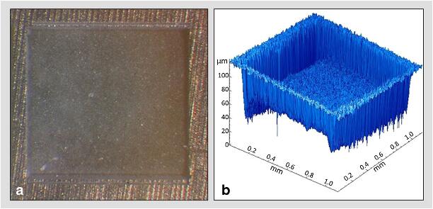
Laser irradiation was performed with an Nd:YVO4laser at1,064 nm (modified Super Rapid, Lumera Laser, Kaisers-lautern, Germany), a pulse duration of 9 ps, and a repetitionrate of 500 kHz. The output power of the laser device wasset to 6 W for all measurements. Laser irradiation time was0.8 s with a delivered total energy of 4.8 J. Employing ascanner system with a scanning speed of 2,000 mm/s (ScanCube 7, Scanlab, Puchheim, Germany), rectangular cavitiesof 1-mm edge length were generated (Fig.1). The scanpattern consisted of individual horizontal lines with a spotsize of 30μm and a spacing of 12.5μm. At the end of eachline, the laser is automatically switched off and moves to thenext line. The laser pulses need to overlap to performhomogeneous ablation patterns. This overlap is determined by scanning speed, repetition rate, and pulse diameter alongeach line, whereas the vertical overlap of two successivelines is affected by pulse diameter and line distance. Param-eters for the present study led to an overlap of 44 % inhorizontal and 84 % in vertical direction. The final cavitywas the result of 12 scans. Prior to temperature measurements,a preliminary survey was performed to assess the ablation ofboth enamel and dentine samples.Employing an optical surfacemeasuring system (MicroSpy, Fries Research and Technology,Bergisch Gladbach, Germany),the ablation depth of 26 speci-mens of each hard tissue was evaluated. Additionally, all sam-ples were photographed employing a light microscope (WildM8, Leica Mikrosysteme, Wetzlar, Germany).
Results
Ablation depthProfilometry showed a median ablation depth of 29μm(min, 9μm; max, 87μm; interquartile range, 16μm) inenamel and 43μm (min, 14μm; max, 80μm; interquartilerange, 35μm) in dentine. Ablation depths were statisticallysignificantly different (p<0.05, Mann–Whitney).Maximum temperature increaseHeat generation could be observed during ablation of bothenamel and dentine specimens. In general, enamel samplesshowed a higher maximum temperature increase than den-tine samples (p<0.05). However, temperature increasedepended on the thickness of the respective material (p<0.05) (Figs.2and3). Lowest delta temperature values couldbe observed in the 3-mm thickness group for dentine (me-dian delta value, 0.7 K; min, 0.5 K; max, 1.2 K) and in the2-mm thickness group for enamel (median delta value,7.3 K; min, 4.9 K; max, 23.9 K). Highest delta temperaturevalues could be observed in the 1-mm thickness group forboth dentine (median delta value, 6.8 K; min, 3.6 K; max,13.6 K) and enamel (median delta value, 44.3 K; min,29.3 K; max, 67.0 K).
dental laser tips


