Pulsed Laser Sintering of Photo-active Calcium Phosphates for Dental Enamel Restoration
Introduction: Acid erosion and wear of natural enamel leading to sensitivity of teeth is a common condition amongst large population worldwide. It is a lifestyle related condition, resulting from the progressive loss of mineral phase from enamel surface. Unlike bone, the dental enamel is a dead tissue and cannot regenerate itself and it must be restored for preventing exposure of softer dentinal tubules and nerves [1], which is the root cause of pain. At present there is no long-term treatment except the daily use of brand tooth pastes, regular use of which provide some protection. Consequently, the dental condition globally is on rise. Novel enamel restoration procedure using lasers: We investigated the laser sintering behaviour of rare-earth oxide doped calcium phosphate suspensions, which was applied on the surface extracted, disinfected, y-ray irradiated teeth, surfaces of which were prepared apriori using dilute phosphoric acid. The natural dental enamel is a hydroxyl apatite (HAp) [2]. We refer to rare-earth doped calcium phosphates as the photo-active enamel mineral (PAEM), which was then irradiated with both pulsed and CW lasers. For effective photo-activation of synthetic mineral leading to sintering with the natural enamel using pulsed laser, the calcium phosphate phase was doped with 1-2 wt% ofYb203 or Er203 oxide, which was dissolved into the suspension together with 2 wt% of aluminium phosphate (AIP04), 2 wt% calcium fluoride (CaF2) and the remaining calcium phosphate constituents, which makes the majority of mineral. The synthesized mineral, unlike natural and synthetic needle like HAp, has platelet structure for providing larger surface occlusion, rapid heat transfer, and large surface area for sintering (Figure la). The rare-earth oxides as dopant facilitate strong energy absorption through resonant band of the RE-oxide, and facilitate sintering during ultra fast (femto-second) and CW laser irradiation. To date we have analyzed and compared sintering of PAEM with 800 nm (Ti sapphire laser, 430 mW, 135 fs, 83 MHz) in Figure Ib, 1520 nm (Cr 4 +-laser, 150 mW, 100fs, 2.5 OHz) in Figure Ic, and 980 nm CW diode laser (500 mW CW) in Figure Id. The sintering was studied as a function of time and it was found that with high repetition rate non-resonan t laser, e.g. at 800 nm, and the resonant 1520 nm, the sintering time was less than 2 minutes. Prolonged irradiation beyond 2 minutes resulted in the structural damage as shown in Figure I b, with surrounding sintered region. The irradiated surfaces were analysed for heat accumulation by undertaking temperature measurements on pressed synthetic mineral pellets, and the maximum temperature rise of 30°C was recorded on small discs of I mm in thickness. The mineral constituents such as CaF2 and AIP04 are incorporated for enhancing acid and wear resistance, and ease of sintering during laser irradiation. As a preliminary step to full in-vitro and in-situ evaluation of laser sintered materials, the hardness of the sintered materials were compared with respect to exposed dentine surface and the natural enamel. The hardness of natural enamel is 3.50Pa [3] compared to 0.25 OPa for an exposed surface with thin and eroded layer of mineral. The micro hardness of laser sintered materials is thickness dependent and 1.20Pa for less than 20 11m sintered layer using 980 nm. The hardness for fs-Iaser sintered materials will be presented at the conference together with the data on acid resistance. Microscopic and X-ray diffraction analysis shows that the synthetic phosphate tends to transform into hydroxyl apatite under laser irradiation. In conclusion, although the nano-second pulsed laser [4] with slow repetition rates have been quite popular together with CO2 lasers for general surgical procedure in dentistry, these lasers are unsuitable for restoration of enamel surface. The ultra-fast high repetition rate lasers of P AEM offer good opportunity for bringing a new application of laser in clinical dentistry for the prevention of acid erosion leading to hypersensitivity .
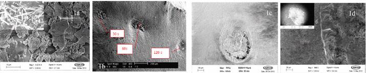
Figure I: a) Platelet phosphate and needle shape HAp (inset), b) microstructure of irradiated minerals with 800 nm 100 fs 80MHz, c) 1520 nm 2.5 GHz, 150 mW power, the focussed laser spot was where there is crack, d) 980 nm CW laser with the laser spot (inset). The rosette-like crystals grow after few minutes while the liquid recesses.


